SACRUM ANATOMY


Sacrum has distinctive anatomical differences from other parts of the spine. This article reviews anatomic properties of the sacrum.
  • Sacrum (Bone)
  • Sacral Ligaments
  • Vascular Anatomy of Sacral Region
  • Sacral Neural Anatomy

Sacrum

The adult sacrum consists of five fused sacral vertebrae. At birth, each vertebral body is separated by an intervertebral disc. The two caudal bodies fuse at approximately the 18th year of life, and the process of fusion continues rostrally until the S1–2 interspace finally fuses by 30 years of age. The development of fused vertebrae is dependent on normal weight bearing. The sacrum is wedge shaped, and the broad base of S1 forms the posterior segment of the pelvic ring. The upper sacral vertebrae articulate with the ilium bilaterally. The ventral sacrum is concave and contains the ventral sacral foramina of the S1–4 nerve roots. Exiting the foramina are the paired sacral nerves and associated vascular elements. The transverse prominences between each pair of foramina represent the area where the intervertebral disc was once located. The pars lateralis (lateral mass) is the area lateral to the ventral foramina. The lateral surfaces of the upper two or three sacral vertebrae form an ear-shaped “auricular” surface, which articulates with the ilium on each side. Ventrally, the lateral masses are marked by neural grooves that run laterally from each of the foramina. The large lateral masses of S1 are known as the alae. On the anterior aspect of each ala is a rounded bony groove formed by the lumbosacral trunk. The dorsal surface of the sacrum is convex and has an irregular surface that includes median, intermediate, and lateral sacral crests representing the fused spinous, articular, and transverse processes, respectively. The shallow grooves between the median and intermediate crests are formed by fused laminae. The dorsal rami of the upper four sacral nerves and associated vascular structures pass through the four pairs of dorsal sacral foramina, located between the intermediate and lateral sacral crests. The dorsal sacral foramina are much smaller than the corresponding ventral sacral foramina. The laminae of the fifth (and on occasion the fourth) vertebra fail to fuse in the midline and form the sacral hiatus, which is the caudal opening of the sacral canal. The sacral cornua, remnants of the inferior articular process, lie on each side of the sacral hiatus.

Sacrum bony anatomy

Sacral Ligaments

Because the lumbosacral and sacroiliac joints transmit the entire weight of the body to the hip bones and lower limbs, these joints and their supporting ligaments must be very strong. The strong dorsal ligamentous complex includes the interosseous ligaments and the dorsal sacroiliac ligaments. The very stout interosseous ligaments connect the sacral tuberosities to the overhanging bone of the iliac tuberosities and represent the single strongest ligaments binding the sacrum to the ilium. The dorsal sacroiliac ligaments are divided into deep (short) and superficial (long) parts. The deeper ligaments connect the sacral and ilial tuberosities and are composed of horizontally oriented
fibers; the more superficial ligaments are oriented vertically and stretch from the posterior superior iliac spine to the tubercles of the lateral sacral crest. The caudal portions of the superficial dorsal sacroiliac ligaments blend with the sacrotuberous ligaments.
The ventral ligamentous complex includes the ventral sacroiliac ligaments and the lumbosacral ligaments. The ventral sacroiliac ligament is a weak fibrous band that attaches to the base and lateral part of the sacrum and to the medial margin of the auricular surface of the ilium.
Three sets of accessory ligaments—the sacrospinous, sacrotuberous, and iliolumbar ligaments—also function to strengthen the pelvic girdle. The iliolumbar ligament originates on the transverse process of L5 and courses caudally and laterally to insert on the ilium. Some fibers stretch ventrally to merge with the ventral sacroiliac ligament as the lumbosacral ligament. The sacrospinous ligament connects the lateral and anterolateral surfaces of the sacrum and coccyx with the ischial spine. This ligament divides the sciatic notch into greater and lesser sciatic foramina. Finally, the sacrotuberous ligament, an extensive structure, originates broadly from the posterior superior iliac spine and the dorsal and lateral aspects of the sacrum and coccyx to form a dense narrow fibrous band that inserts on the ischial tuberosity.

Sacrum ligaments

Vascular Anatomy of Sacral Region


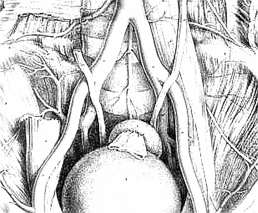
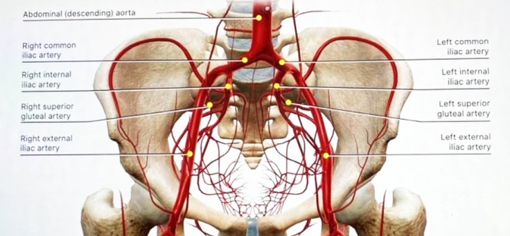
The abdominal aorta bifurcates at the L4 level. The small median or middle sacral artery arises from the posterior surface of the abdominal aorta close to the bifurcation and descends vertically along the pelvic surface of the sacrum. It gives rise to several small parietal branches that anastomose with the lateral sacral arteries and to small visceral branches that anastomose with the superior and middle rectal arteries.
The aorta divides into the common iliac arteries, which travel laterally and inferiorly. At approximately the L5–S1 disc space, the common iliac arteries divide into the external and internal iliac arteries. The ureter crosses the external iliac artery anteriorly. The internal iliac artery is separated from the sacroiliac joint by the internal iliac vein and the lumbosacral trunk.
The iliolumbar artery can arise from the common iliac artery, although it more commonly is the first branch of the internal iliac artery. This vessel runs superomedially, passing anterior to the sacroiliac joint and posterior to the psoas muscle. It later turns laterally and upward to divide in the region of the iliac fossa. The lateral sacral artery is usually the second branch of the internal iliac artery, although it can originate from the superior gluteal artery. These vessels, usually a superior one and an inferior one, sometimes arise from a common trunk. They pass medially and descend downward anterior to the sacral ventral rami, giving branches that enter the ventral sacral foramina to supply the spinal meninges and the roots of the sacral nerves. Some branches pass from the sacral canal through the dorsal foramina to supply the muscles and skin overlying the sacrum. Most tumors arising from the sacrum or presacral space, as well as some intraspinal masses, derive at least a part of their blood supply from the medial and lateral sacral arteries. Enlargement, displacement, or encasement of these vessels may be seen on angiography.
Abdominal vessels
The next two branches of the internal iliac artery are the superior and inferior gluteal arteries. The superior gluteal is a large artery that passes anteriorly across the lumbosacral trunk as the trunk passes over the ala. It then turns posteriorly between the lumbosacral trunk and the ventral ramus of the first sacral nerve to leave the pelvis through the superior part of the greater sciatic foramen, superior to the piriformis muscle. The inferior gluteal artery passes posteriorly to pierce the sacral plexus more inferiorly (most often between S2 and S3) and exits the pelvis through the inferior part of the greater sciatic foramen, inferior to the piriformis muscle.
The venous anatomy of the region generally parallels that of the arterial anatomy, but it is more variable. There are several important features of the venous anatomy to note. First, the vena cava lies to the right of the aorta at the bifurcation of the common iliac vessels. The right common iliac vein passes posterior to the common iliac artery, and it is therefore shorter than the left common iliac vein. Second, the middle sacral vein, which is occasionally doubled, drains into the left common iliac vein rather than directly into the inferior vena cava. Finally, the iliolumbar veins drain into the common iliac veins rather than into the internal iliac veins.


Sacral Neural Anatomy

The thecal sac ends blindly at the S2 level. The lower sacral and coccygeal nerves emerge from the sac, as does the extradural portion of the filum terminale. The upper four roots exit the sacrum through the paired ventral and dorsal sacral foramina. The fifth sacral roots, the coccygeal roots, and the filum exit the sacrum caudally through the sacral hiatus. The filum terminale extends to its point of fusion with the periosteum of the first coccygeal segment.
The sacral plexus is formed by the ventral rami of six roots: L4 through S3 and the upper part of S4. The lumbosacral trunk (the conjoined L4–5 roots) crosses the anterior aspect of the ala of the sacrum, descends obliquely in front of the sacroiliac joint, and enters the pelvis deep to the parietal pelvic fascia. It crosses the superior gluteal vessels and joins the first sacral root. The sacral plexus is located on the anterior surface of the piriformis muscle, deep to the parietal (Waldeyer’s) fascia. Except for the nerves to the piriformis muscle, the perforating cutaneous nerves, and the nerves to the pelvic diaphragm, essentially all branches of the sacral plexus leave the pelvis through the greater sciatic foramen.
The most important derivatives of the sacral plexus are the sciatic and pudendal nerves. The latter is unique in exiting the greater sciatic foramen only to reenter the lesser sciatic foramen by hooking around the sacrospinous ligament. It supplies the muscles of the perineum, including the external anal sphincter, and provides sensory information to the external genitalia.
The coccygeal plexus is derived from the ventral rami of S4 and S5 as well as the coccygeal roots. It lies on the pelvic surface of the coccygeus muscle. It innervates the coccygeus muscle and provides some perianal sensation.
Both the sympathetic and parasympathetic components of the autonomic nervous system have an intimate relationship with the sacrum. The sacral sympathetic trunk, continuous with the lumbar sympathetic trunk, descends against the ventral surface of the sacrum, converging in front of the coccyx to form the unpaired ganglion impar. Three or four sacral trunk ganglionic enlargements are found on each side of the midline, just medial to the ventral sacral foramina. No white rami communicantes are present in this region; however, the postsynaptic gray rami communicantes from each ganglion join the corresponding sacral or coccygeal nerves for distribution to sweat glands, blood vessels, and erector pilori muscles. In addition, the sacral sympathetic trunks provide fine branches to the superior hypogastric plexus. The superior hypogastric plexus is the caudal continuation of the periaortic sympathetic plexus; it lies on the anterior surface of the fifth lumbar vertebra and upper sacrum in the retroperitoneal tissue. Fibers of the superior hypogastric plexus diverge into right and left hypogastric nerves opposite the first sacral vertebra. The term hypogastric nerve may be a misnomer, because the structure is really a narrow plexus of fibers. The hypogastric nerves represent the principal sympathetic inputs to the inferior hypogastric plexus. The parasympathetic contributions to the pelvic plexi arise from the ventral S2 to S4 nerve roots. These preganglionic fibers form the pelvic splanchnic nerves (nervi erigentes). The parasympathetic system provides motor innervation to the detrusor muscle of the bladder and is primarily responsible for the vascular reflexes that sustain erectile function. The sympathetic system plays less of a role in normal voiding reflexes but is important for male fertility by promoting timely transport of spermatozoa from the testes to the seminal vesicles and by coordinating reflexes responsible for ejaculation.


Any comment about this page?
Your feedback is appreciated. Please click here.

Follow & Share Scientific Spine  Bookmark and Share Subscribe

To join Scientific Spine mailing list, click here.

You are here: Scientific Spine > Spinal Anatomy > Sacrum Anatomy