 |
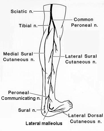
Anatomy of the Sural Nerve There are two sural nerves: medial and lateral sural cuteus nerves (see picture below). Medial sural cutaneus nerve borns from the tibial nerve just below knee joint. It runs downward across between the heads of gastronemius. Lateral sural cutaneous nerve arises from common peroneal nerve above knee joint and runs down posterolateral aspect of calf. Medial and lateral sural cutaneus nerves are connected by peroneal communicating branch. Thus, the union of these 3 nerves constitutes the sural nerve. Sural nerve passes down posterolateral side of leg & onto dorsal aspect of lateral side of foot, giving rise to lateral calcaneal branches (medial branch supplied by tibial nerve). The nerve runs with the small saphenous vein on the posterior leg just lateral to the achilles tendon, and its terminal branches consist of lateral dorsal cutaneous nerve and the lateral calcaneal branches. |
References
Coert JH, Dellon AL. Clinical implications of the surgical anatomy of the sural nerve. Plast Reconstr Surg. 1994;94:850-855.
Any comment about this page?
Your feedback is appreciated. Please click
here.
To join Scientific Spine mailing list, click here.